Positron Emission Tomography (PET) combined with Computed Tomography (CT), or PET CT imaging, is an advanced diagnostic tool that provides crucial information about the body’s functions and structures. Used extensively in the detection of cancer, cardiovascular diseases, and neurological disorders, PET CT combines the benefits of two imaging technologies to deliver detailed, precise insights that guide healthcare providers in making informed decisions. In this blog, we’ll explore the science behind PET CT imaging, what patients can expect from the procedure, and the exciting future developments in PET CT technology.
How PET CT Works
PET CT imaging merges two powerful technologies — Positron Emission Tomography (PET) and Computed Tomography (CT) — to offer a comprehensive view of the body. Here’s how each part contributes:
- PET (Positron Emission Tomography): PET scanning detects the metabolic activity in tissues by using a small amount of a radioactive tracer injected into the body. These tracers emit positrons (tiny particles) that are detected by the scanner, providing detailed information about the activity and function of organs and tissues. This is particularly useful for detecting cancer cells, as tumors tend to have a higher metabolic rate than normal tissues.
- CT (Computed Tomography): CT scanning creates detailed images of the body’s internal structures, such as bones, organs, and blood vessels. Using X-rays, the CT scan produces cross-sectional images that provide a detailed view of the body’s anatomy. This helps physicians localize areas of concern detected by the PET scan and provides additional information on size, shape, and exact location of potential abnormalities.
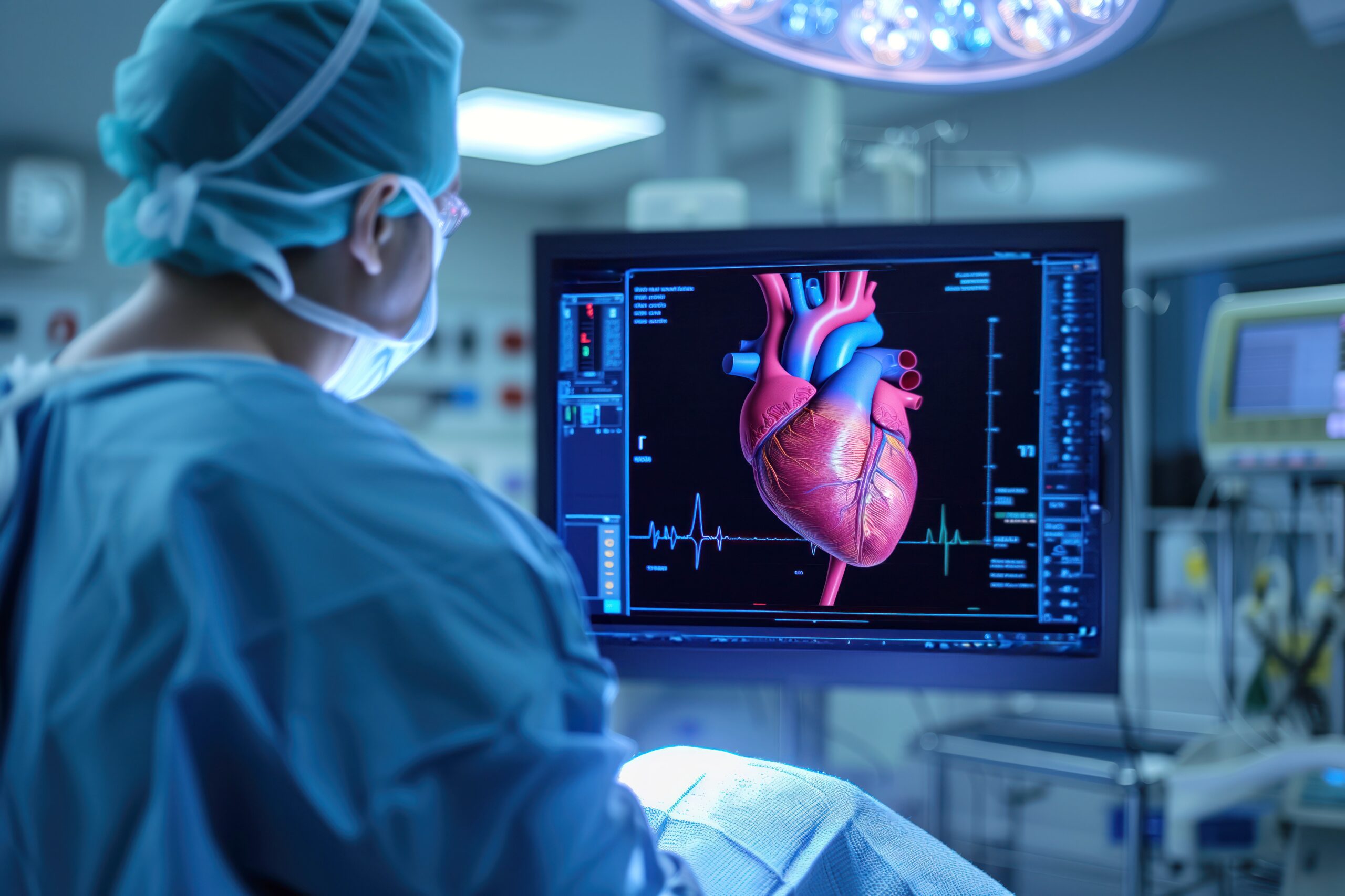
When combined, PET CT imaging provides both functional and anatomical information in one scan, offering a more accurate, detailed picture than either technology could provide on its own. It’s a valuable tool for diagnosing, staging, and monitoring treatment progress for conditions such as cancer, heart disease, and brain disorders.
What to Expect from the Scan
If you’re preparing for a PET CT scan at a facility like PET CT of Miami, here’s what you can expect during the procedure:
- Pre-scan Preparation: Before your PET CT scan, you may be asked to fast for a few hours. This is to ensure that the tracer will be absorbed more efficiently by your body. Depending on the area being scanned, you might be asked to drink water or follow specific instructions.
- Injection of the Tracer: A small amount of a radioactive tracer will be injected into your vein. This tracer will travel through your bloodstream and be absorbed by the tissues in your body. Depending on the area of concern, the tracer will concentrate in different regions, allowing the scanner to identify areas of heightened activity.
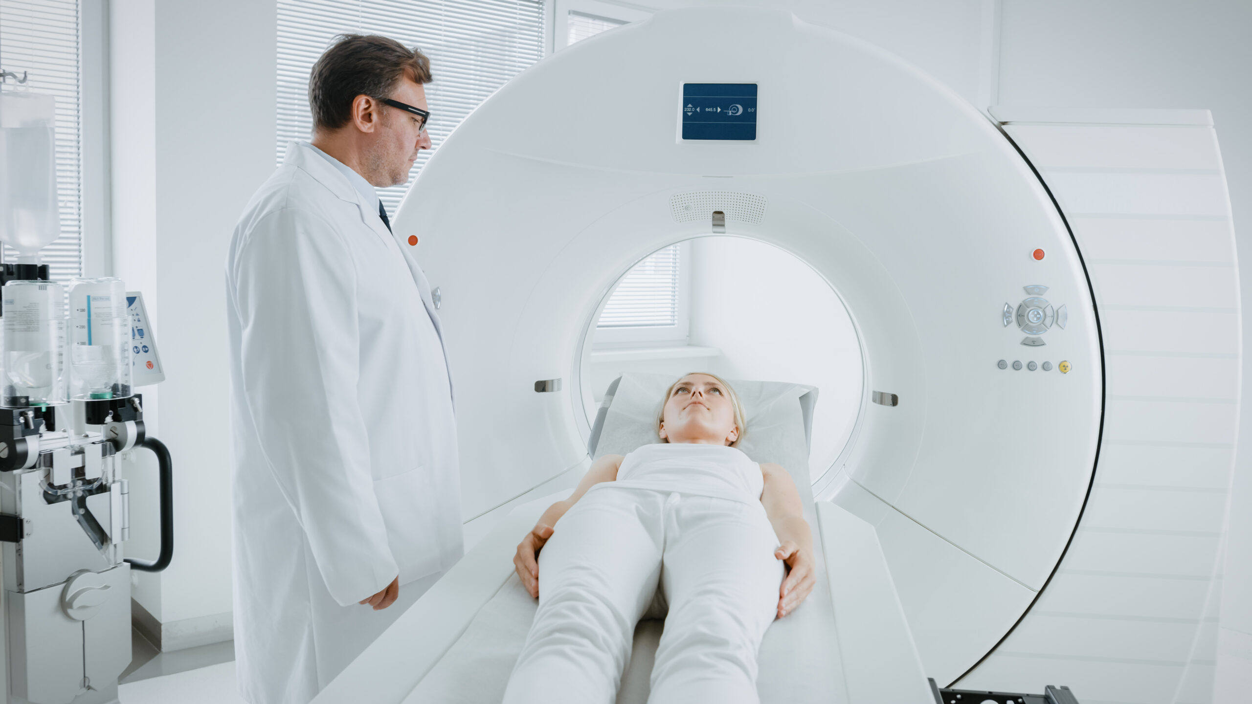
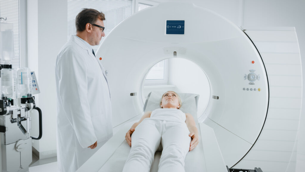
- Scan Procedure: Once the tracer has had time to work (typically 30 to 60 minutes), you will be positioned on the PET CT scanner. During the scan, you’ll need to lie still while the scanner takes images of your body. The procedure is non-invasive, although you may experience some mild discomfort from lying still or the injection.
- Duration: The entire procedure usually lasts between 30 minutes to an hour. You can expect to return to your normal activities shortly after the scan is completed.
While the PET CT scan itself is painless, the preparation and the slight discomfort from the injection may vary from patient to patient. It’s always best to follow your medical provider’s guidelines to ensure an optimal scan.
Future of PET CT Technology
As healthcare continues to evolve, so does the technology behind PET CT imaging. Here are some exciting trends and advancements on the horizon:
- Improved Resolution and Sensitivity: Future developments are focused on increasing the resolution and sensitivity of PET CT scanners. This will allow for earlier detection of diseases and more accurate monitoring of treatment progress, particularly in the fight against cancer.
- Integration with Other Technologies: Researchers are working on integrating PET CT with other imaging technologies, such as MRI and Ultrasound, to create more comprehensive diagnostic tools. This combination can potentially provide more detailed images of the body’s structure and function, further enhancing diagnostic accuracy.
- Reduced Radiation Exposure: Advancements in scanner design and radiopharmaceuticals are helping to reduce the amount of radiation patients are exposed to during a PET CT scan. This is crucial for improving patient safety, especially for those who may need frequent imaging.
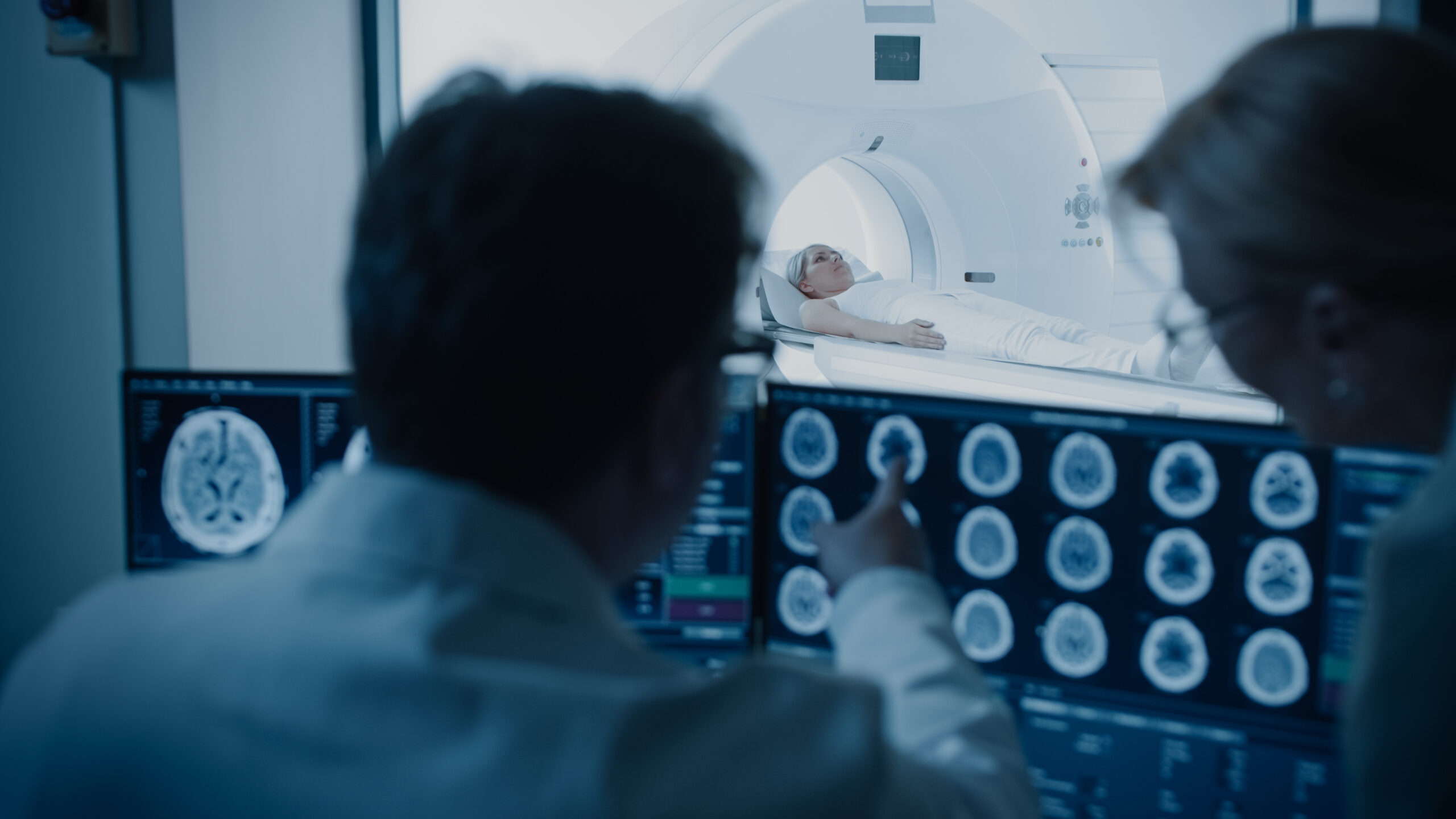
- Artificial Intelligence (AI) and Machine Learning: AI and machine learning algorithms are being integrated into PET CT systems to assist in the interpretation of images. These technologies help radiologists detect anomalies more efficiently and accurately, streamlining the diagnostic process.
As these innovations continue, the future of PET CT imaging looks promising, offering even more precise, personalized, and efficient care for patients.
Conclusion
PET CT imaging plays a vital role in modern diagnostics, providing invaluable insights into the function and structure of the body. By combining the strengths of Positron Emission Tomography and Computed Tomography, it allows doctors to detect and diagnose a wide range of conditions with greater accuracy. Whether you are undergoing a PET CT scan for cancer screening, neurological issues, or cardiovascular disease, you can trust that this advanced technology will provide a comprehensive view of your health. With continuous advancements in PET CT technology, the future of medical imaging is brighter than ever.
Schedule Your Appointment at PET CT of Miami
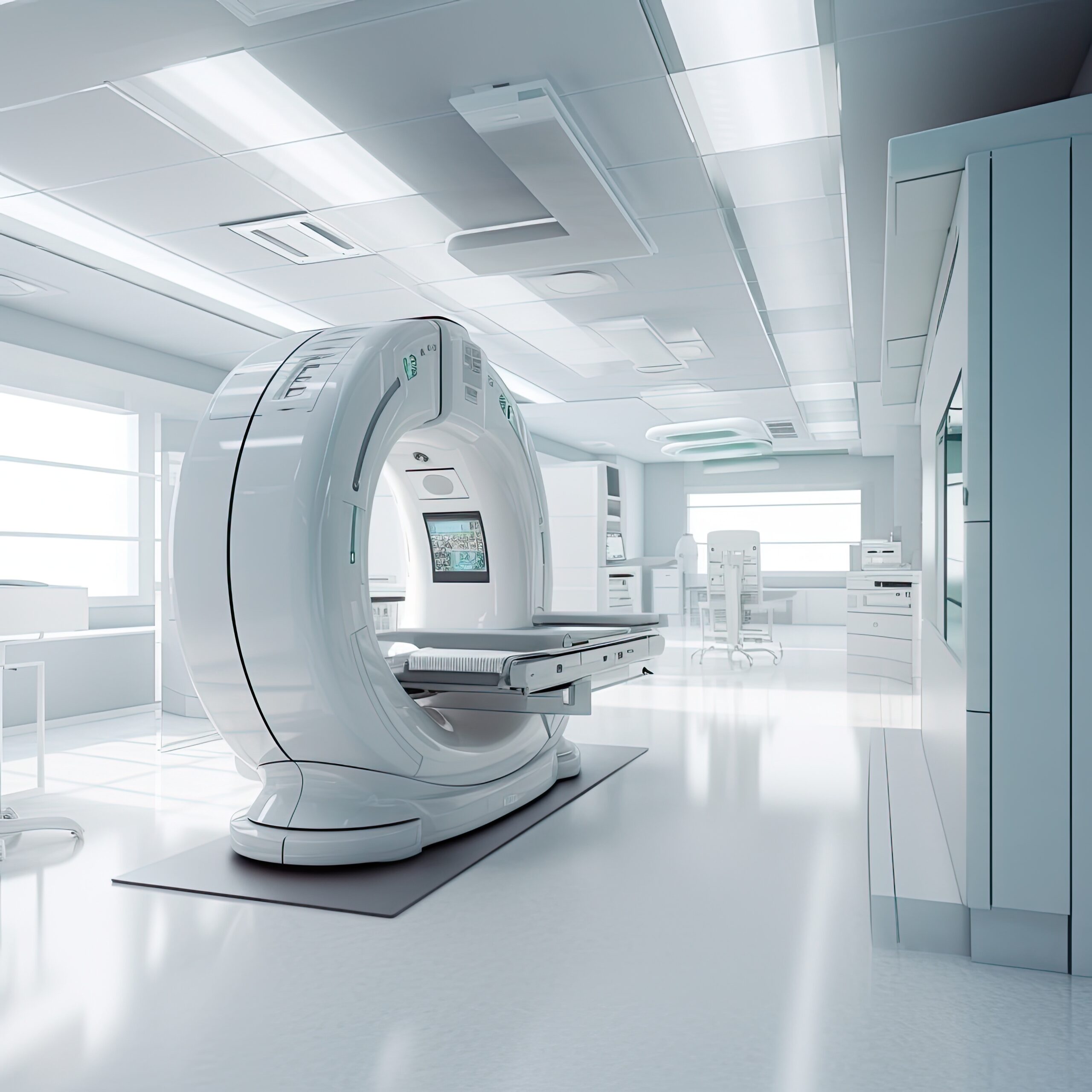
At PET CT of Miami, we specialize in PET CT scans and other diagnostic imaging services to provide accurate and timely results. Our state-of-the-art equipment and expert staff ensure that you receive the highest quality care. Scheduling your appointment is easy — simply visit our website and book your appointment online today!

