Looking inside the brain before symptoms take hold.
The Challenge of Detecting Alzheimer’s Disease Early
Alzheimer’s disease is a progressive neurodegenerative disorder that affects memory, thinking, and behavior. It’s the most common form of dementia, impacting over 6 million Americans—a number projected to double by 2050. Yet, one of the biggest challenges in treating Alzheimer’s is early detection.
By the time memory loss and confusion become obvious, significant damage has already occurred in the brain. That’s why researchers and neurologists now rely on advanced imaging—especially Positron Emission Tomography (PET) scans—to detect Alzheimer’s before outward symptoms appear.
In this blog, we’ll explore how PET imaging works, what it reveals about Alzheimer’s disease, and why it’s becoming a critical tool in early diagnosis and treatment planning.
Understanding the Biological Basis of Alzheimer’s
Alzheimer’s is marked by two key pathological changes in the brain:
- Beta-amyloid plaques – Abnormal clumps of protein that disrupt cell communication
- Tau tangles – Twisted strands of protein inside neurons that block nutrient transport
These changes begin years—even decades—before cognitive decline is noticeable. During this long “preclinical” phase, the brain is silently changing. PET imaging offers a way to see those changes in real-time, long before a diagnosis based on symptoms alone is possible.
What Is a PET Scan, and How Does It Work?
Positron Emission Tomography (PET) is a non-invasive imaging technique that allows doctors to observe metabolic activity or molecular changes inside the brain. It uses a small dose of a radioactive tracer injected into the bloodstream. This tracer accumulates in targeted areas of the brain and emits signals that the scanner converts into detailed 3D images.
In Alzheimer’s care, PET imaging typically uses two types of tracers:
1. FDG-PET (Fluorodeoxyglucose PET)
- Measures how brain cells use glucose (sugar), the brain’s primary fuel
- In Alzheimer’s, certain brain regions (especially in the temporal and parietal lobes) show reduced glucose metabolism
- Useful for distinguishing Alzheimer’s from other types of dementia
2. Amyloid PET
- Uses tracers that bind specifically to beta-amyloid plaques
- Can detect abnormal amyloid buildup even before symptoms appear
- Helps confirm or rule out Alzheimer’s in patients with mild cognitive impairment (MCI)
Some advanced centers also offer Tau PET, which images the second major pathology of Alzheimer’s.
Why Early Detection Matters
Many people assume that there’s no benefit to an early Alzheimer’s diagnosis—but the truth is the opposite. Here’s why detecting the disease in its earliest stage can be life-changing:
- Treatment access: Medications like Lecanemab and Donanemab are designed to target amyloid plaques in the early stages—when they’re most effective
- Lifestyle intervention: Evidence shows that changes in diet, exercise, sleep, and cognitive stimulation can slow progression
- Financial & care planning: Patients and families can prepare while the individual is still able to participate in decisions
- Clinical trials: Many trials now require confirmed amyloid presence via PET imaging for participation
In short, knowledge is power, and PET imaging makes that knowledge possible—often years ahead of traditional tools.
How PET Imaging Differs from MRI or CT in Alzheimer’s Diagnosis
Many patients ask: Why can’t a regular MRI or CT scan detect Alzheimer’s?
Here’s a breakdown:
| Imaging Type | What It Shows | Limitations in Alzheimer’s |
| CT Scan | Basic brain anatomy | Only shows late-stage brain shrinkage |
| MRI | High-resolution brain structure | Can show atrophy but not molecular changes |
| PET Scan | Metabolism and molecular changes | Detects early-stage abnormalities |
PET doesn’t just show brain structure—it shows how the brain is functioning, which is crucial in Alzheimer’s. It reveals the problem before brain cells have visibly deteriorated.
When Should You Consider a PET Scan for Alzheimer’s?
PET imaging isn’t used for routine memory complaints. It’s typically recommended when:
- A patient has mild cognitive impairment (MCI) with unclear cause
- There’s early memory loss with a family history of Alzheimer’s
- The diagnosis is uncertain between Alzheimer’s and other dementias (like frontotemporal dementia or Lewy body disease)
- Confirmation of amyloid plaques is needed for treatment eligibility or clinical trial enrollment
- A person wants proactive screening due to genetic risk (such as APOE4 carriers)
If you’re unsure whether a PET scan is appropriate, your neurologist or primary care doctor can help guide the decision.
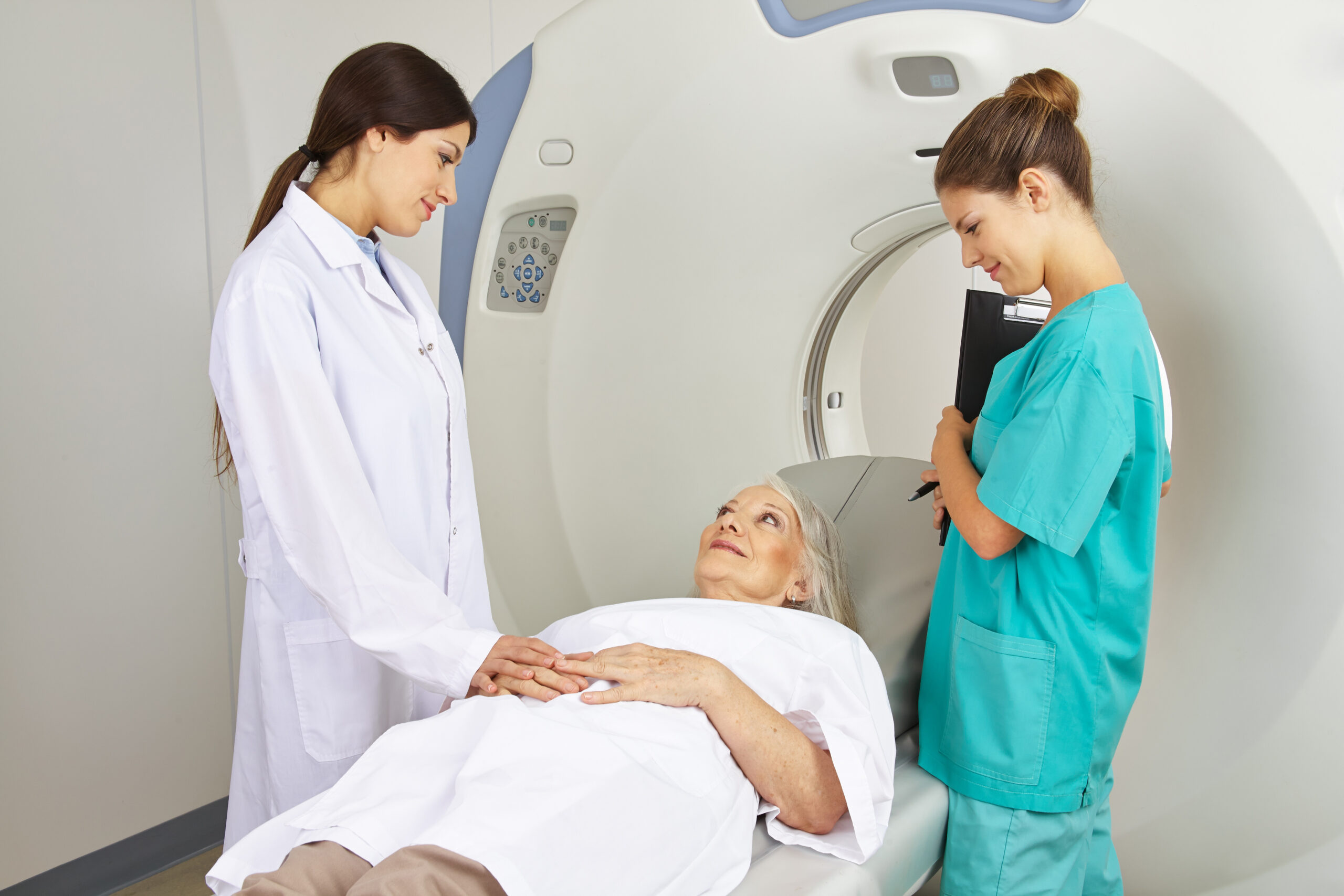
What to Expect During a Brain PET Scan
PET imaging is safe, non-invasive, and relatively comfortable. Here’s a step-by-step overview:
Before the Scan:
- You’ll be asked to fast for several hours (usually 4–6)
- Avoid caffeine, alcohol, and vigorous exercise beforehand
- Notify your doctor if you’re pregnant or breastfeeding
During the Scan:
- A small IV is placed in your arm for the tracer injection
- You’ll rest quietly for 30–60 minutes while the tracer circulates
- You’ll lie still on a scanning table while the PET scan is performed (about 20–30 minutes)
- The entire process takes about 90 minutes
There is no pain, and side effects are rare. The small amount of radiation used is considered safe and is eliminated naturally within 24 hours.
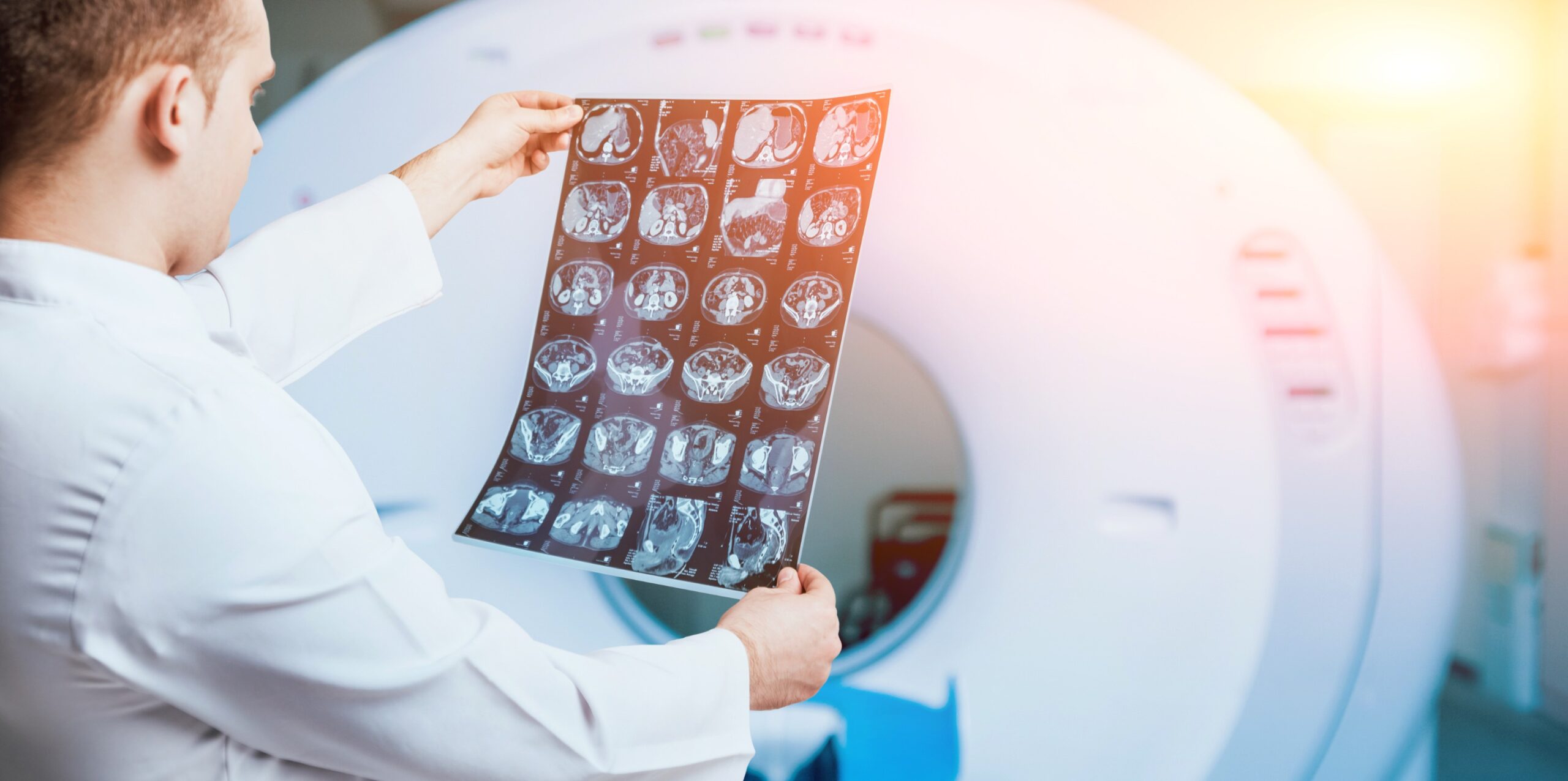
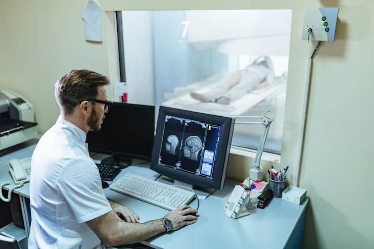
Interpreting the Results
After the scan, a radiologist and/or nuclear medicine specialist will review the images and send a report to your doctor.
What they’ll look for:
- Areas of hypometabolism (reduced activity) in the parietal and temporal lobes
- Signs of widespread or focal amyloid plaque accumulation
- Metabolic patterns that distinguish Alzheimer’s from other forms of dementia
Your doctor will review these findings with you and explain:
- Whether Alzheimer’s is likely or unlikely
- If additional testing is needed
- What the next steps are in treatment or lifestyle planning
Why Choose PET CT and MRI of Miami for Brain PET Imaging?
We are proud to be one of South Florida’s trusted providers of brain PET imaging for cognitive and neurological evaluation. Our commitment to accuracy, comfort, and compassionate care includes:
- State-of-the-art PET/CT technology
- Fast scheduling—often within a week
- Amyloid and FDG PET options available
- Board-certified neuroimaging specialists
A calm, patient-first environment with full pre-scan support - Seamless coordination with your neurologist or primary doctor
We believe that early answers lead to better outcomes—and we’re here to provide both.
Take the First Step Toward Clarity
If you or someone you love is experiencing memory problems, confusion, or early cognitive symptoms, don’t wait. A brain PET scan could provide clarity, confidence, and a path forward.📍 PET CT and MRI of Miami
📞 Call us at (305) 229-2020
🌐 Schedule at petctofmiami.com