How magnetic imaging helps identify seizure causes and guide treatment.
Epilepsy Isn’t Just About Seizures—It’s About Answers
For the 65 million people worldwide living with epilepsy, the journey to diagnosis and management often begins with one essential tool: imaging. Among all available options, Magnetic Resonance Imaging (MRI) remains the gold standard for understanding what’s happening inside the brain—and why seizures may be occurring.
If you or a loved one has recently been diagnosed with epilepsy, or you’re experiencing unexplained seizures, this guide will walk you through how MRI is used in epilepsy diagnosis and treatment planning, and why it’s often the first—and most important—step toward personalized care.
Why MRI Matters in Epilepsy
Epilepsy is a neurological disorder characterized by recurring, unprovoked seizures. While some forms of epilepsy are genetic or idiopathic (with no identifiable cause), many are the result of structural abnormalities in the brain—and MRI is uniquely suited to detect those.
Unlike CT scans, which are better for acute emergencies like trauma or bleeding, MRI provides high-resolution images of soft brain tissue, allowing doctors to see fine details like:
- Scar tissue from past injury
- Tumors or cysts
- Cortical malformations (congenital anomalies)
- Signs of stroke, infection, or inflammation
- Areas of atrophy or degeneration
In short: MRI doesn’t just show if something is there—it shows exactly where, how big, and how it affects surrounding tissue.
What Doctors Are Looking For on an Epilepsy MRI
MRI scans for epilepsy are performed using a dedicated epilepsy protocol, which includes:
- Thin slice images (1–3 mm resolution)
- Specific sequences (T1, T2, FLAIR, diffusion-weighted)
- Imaging of the temporal lobes, hippocampus, and other seizure-prone regions
- Sometimes with contrast to enhance detail
Common findings include:
- Mesial Temporal Sclerosis (MTS): Shrinking or scarring of the inner temporal lobe, often associated with temporal lobe epilepsy
- Focal Cortical Dysplasia (FCD): A malformation of cortical development, frequently found in children with epilepsy
- Tumors or Vascular Malformations: May irritate nearby brain tissue, triggering seizures
- Hippocampal Atrophy: Often correlates with memory issues and seizure activity
Even if no abnormalities are seen (a “normal MRI”), the scan is still valuable. It helps rule out other causes and may lead to additional testing, like functional MRI, PET, or EEG monitoring.
When Is MRI Used in the Epilepsy Workup?
MRI is usually ordered early in the diagnostic process after a seizure has occurred—especially if the patient:
- Has never had a seizure before
- Has focal (partial) seizures that affect one part of the body
- Shows neurological changes (e.g., speech, vision, or coordination issues)
- Has developmental delays or head trauma history
- Is being evaluated for surgery
According to the International League Against Epilepsy (ILAE), all patients with focal epilepsy or suspected structural abnormalities should undergo high-quality MRI as part of the standard evaluation.
MRI’s Role in Surgical Planning for Epilepsy
For people with drug-resistant epilepsy—meaning seizures persist despite trying 2 or more medications—surgery may be an option. But for surgery to succeed, doctors must know exactly where the seizures are coming from and whether the source can be removed safely.
MRI plays a crucial role in:
- Localizing seizure onset zones (e.g., left temporal lobe)
- Mapping the relationship of abnormal areas to vital structures (language, memory, movement)
- Assessing whether lesions can be resected without impairing function
- Guiding stereotactic EEG electrode placement if deeper monitoring is needed
- Creating surgical roadmaps for neurosurgeons
In some centers, MRI is combined with fMRI (functional MRI) to identify regions of brain function—particularly useful when planning resections in or near “eloquent” brain areas.
What If Your MRI Is Normal?
Up to 30–40% of people with epilepsy may have a normal MRI, especially in generalized or genetic epilepsy types.
This doesn’t mean your epilepsy isn’t real—it simply means the structural cause isn’t visible with current imaging. In such cases, your doctor may explore:
- Functional imaging like PET or SPECT
- Long-term EEG monitoring
- Genetic testing or metabolic evaluations
- Repeating MRI with higher resolution or newer protocols
Sometimes, repeat MRI years later reveals changes that weren’t visible before. Technology is constantly evolving—and so is your care plan.
What to Expect During an Epilepsy MRI
Preparation:
- Wear loose-fitting clothes without metal
- Remove all jewelry, piercings, or metallic accessories
- Inform your technologist if you have implants or devices
- No fasting is usually required unless contrast is planned
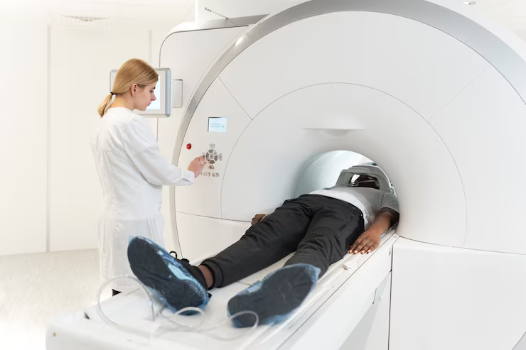
During the Scan:
- You’ll lie on a padded table that slides into the scanner
- The MRI is loud—you’ll receive earplugs or headphones
- You must remain still for 30–45 minutes for optimal image quality
- If contrast is needed, a small IV injection will be given mid-scan
For patients with claustrophobia or sensory issues, we offer:
- Open MRI or wide-bore machines
- Calming music and visual guides
- Sedation options (with prior coordination)
How MRI Fits Into Your Full Epilepsy Treatment Plan
MRI is just one part of a comprehensive epilepsy evaluation. Depending on your symptoms and response to treatment, your care team may also use:
- EEG monitoring to capture electrical activity
- PET scans to assess brain metabolism
- Neuropsychological testing for memory, focus, and language
- Medication trials or device therapy (like VNS)
MRI findings often direct the course of treatment, helping your neurologist determine:
- Which medications may work best
- Whether surgery or other interventions are safe
- How your condition may progress over time
Why Choose PET CT and MRI of Miami for Epilepsy Imaging?
At PET CT and MRI of Miami, we understand that imaging for epilepsy isn’t one-size-fits-all. That’s why we offer:
- Epilepsy-specific MRI protocols
- State-of-the-art high-field and open MRI scanners
- Fast appointments, including same-day slots
- Board-certified radiologists with neuroimaging expertise
- Comfort-first approach for pediatric and adult patients
Whether you’re early in your diagnostic journey or preparing for surgery, our imaging center is here to support you with answers, accuracy, and empathy.
Ready to Schedule Your Epilepsy MRI?
If you or a loved one has been experiencing seizures or unexplained neurological symptoms, don’t wait. An MRI can provide the critical clarity needed to move forward with confidence.📞 Call us at (305) 229-2020
🌐 Visit petctofmiami.com
📍 Located in the heart of Miami, serving South Florida with care and precision